Virginia Equine Imaging
A. Kent Allen, D.V.M. and Associates
Patient
- 16 yo, Thoroughbred, Gelding used as an eventing horse.

History
- Patient presented for an acute onset of severe LF lameness on 7/20/23. Evaluation revealed moderate to severe sensitivity over the medial heel and medial quarter of the LF foot.
- Abscess was drained from medial sole area.
- The horse was sent home with instructions of soaking the foot and applying poultice bandages to help draw out the abscess.

Diagnosis
Radiography
There was a moderate periosteal reaction at the dorsal aspect of the second phalanx. This finding was new when comparing with X-rays taken 2 months before.
 |
 |
Diagnosis
Ultrasound
There was moderate enlargement of the proximal aspect of the medial collateral ligament of the distal interphalangeal joint.

Diagnosis
- The horse was referred to a surgical center for further diagnosis and treatment of the severe lameness. During hospitalization, a LF coffin joint tap was performed and the results were within normal limits. Advanced diagnostic imaging (CT) revealed an enthesopathy of the collateral ligament.
- Because of the collateral ligament involvement it was decided to perform a MRI through VEI.

Diagnosis
Magnetic Resonance Imaging
- Medial collateral distal interphalangeal (DIP) ligament disruption in the distal half.
- Bone edema at the distal insertion of the medial collateral ligament, as well as throughout the quarter.
- Severe distension of the coffin joint.
- Moderate distension of the navicular bursa.
- Sclerosis of the coffin bone at the lateral and medial distal collateral fossa.

Treatment plan:
- LF medial collateral ligament shoe with double clips.
- LF coffin joint injection with PRP.
- US guided injection of the LF medial collateral ligament with PRP.
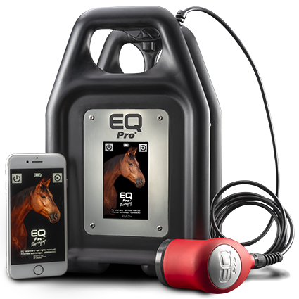
- EQ Pro Therapy on the LF medial collateral ligament 2-3 times per week.
- 4 weeks of stall rest.
EQ Pro Therapy:
- The horse received 2-3 Low-Frequency Therapeutic Ultrasound sessions per week for 3 months.
- A total of 23 treatment sessions were applied using the following protocol:
The Gray Transducer was used first for 5 minutes at 65% power output and then the Blue Transducer was used for 10 minutes at 65% power output on the LF medial collateral ligament of the coffin joint inside of the hoof wall.

Progress
- A lameness evaluation was performed 30 days after the diagnosis and the horse was sound at a walk.
- It was recommended to start with 5 minute hand-walks and 20 minutes of turnout in a small pen once daily.
- He was started on Trazodone to try to keep him quiet during the rehabilitation period.

Progress
- Injections of the LF coffin joint and LF medial collateral ligament were performed 2 months after the diagnosis.
- During the lameness exam performed a month later, the horse presented a 1/5 LF lameness on the straight away trot.
- It was recommended to increase the turnout time to 30-45 minutes once a day.
 Radiograph guided injection of the medial collateral ligament.
Outcome:
- On lameness evaluation, 4 months after the diagnosis, the horse was sound on the straight away trot and lunging in both directions.
Outcome:
- The horse was gradually returned to work with the objective of using him as a trail horse.
- During his most recent lameness examination (9/20/24), the horse was sound at a trot and the owner reports that he has been performing as expected.

Conclusion
- In our clinical experience, the use of Low-Frequency Ultrasound Therapy has demonstrated significant benefits in treating horses with collateral ligament injuries.
- Horses treated with Low-Frequency Ultrasound Therapy have shown improved outcomes compared to those treated with orthobiologics alone.
- The combination of Low-Frequency Ultrasound and Orthobiologic Therapy in our clinic, has been associated with:
.
– Shorter recovery periods.
– Enhanced healing of the collateral ligament.
Conclusion
- Based on our cases, we believe that the use of Low-Frequency Ultrasound Therapy offers a valuable addition to traditional treatments, providing an effective and non-invasive option for managing collateral ligament injuries in horses.


