Low Frequency Ultrasound Therapy Treatment of Superficial Digital Flexor Tendon

Barbara Riccio
DVM, PhD, ISELP cert, DACVSMR, DECVSMR
16th WEVA Congress – Verona, ItalyOctober 3-5, 2019

weva logo
BR logo
university of turin logo

2003 Dressage Holsteiner Gelding

  • History: chronic (more than 1 year old) tendinopathy at the right hindlimb superficial digital flexor tendon (RH SDFT)
  • Poor performance and mild RH lameness
  • Degree of RH lameness increased after last competition (March 19, 2017)
  • Competition level: St. George – Intermédiaire
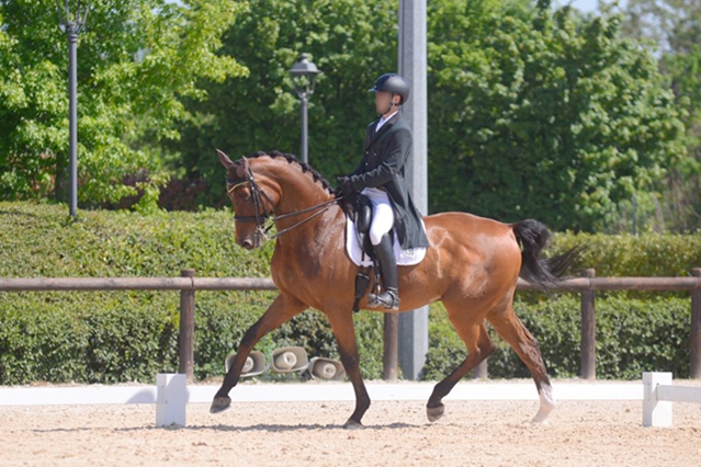
dressage holsteiner gelding

1st examination – March 30, 2017

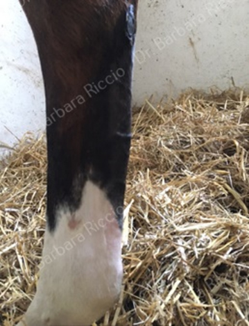
  • Grade 2/5 RH lameness
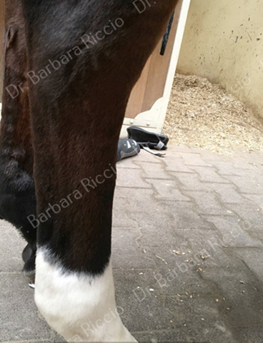
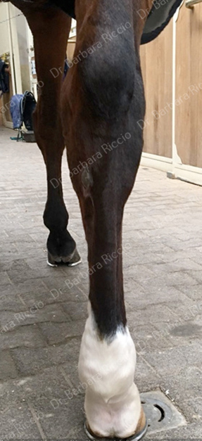
  • Enlarged, hot and painful RH superficial digital flexor tendon
  • Convex plantar profile of the RH tendon region
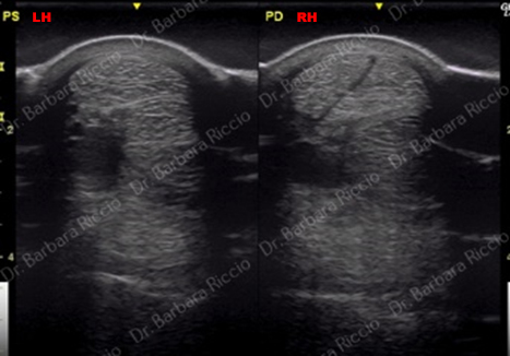
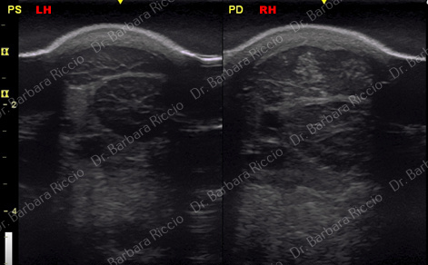
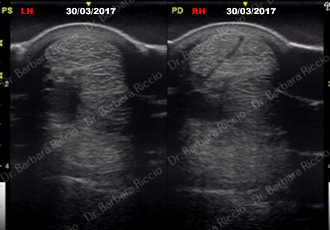
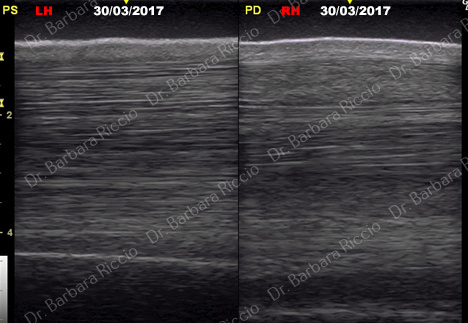
  • 1st ultrasound examination
enlarged hot and painful right hind superficial digital flexor tendon-1
enlarged hot and painful right hind superficial digital flexor tendon-2
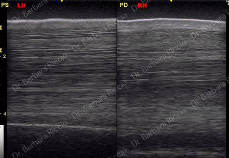
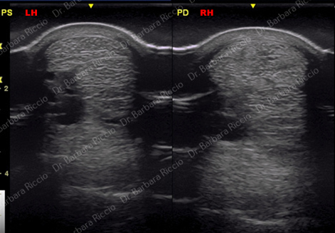
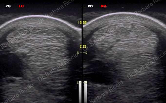
  • RH SDFT increased in size in all scans. Clear split inside the SDFT
  • Using a negative image, the scar inside the SDFT is highlighted
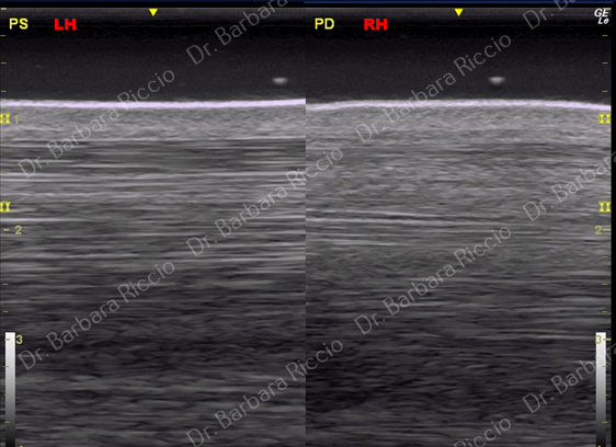
  • Longitudinal scan shows convex profile and short fibers
  • All ultrasonographic signs suggest a chronic tendonitis
equine chronic tendonitis 1
equine chronic tendonitis 2
equine chronic tendonitis 3
equine chronic tendonitis 4

1st examination March 30, 2017 : diagnosis and management

  • Recurrence of RH SDFT tendinopathy
  • Hospitalization 1 month in box with small paddock
  • Walker: 30 minutes twice/day
  • Shoes: aluminium large toe and narrow branches
  • 10 low frequency ultrasound therapy treatments (3 per week for 2 weeks, then 2 per week for 2 weeks)
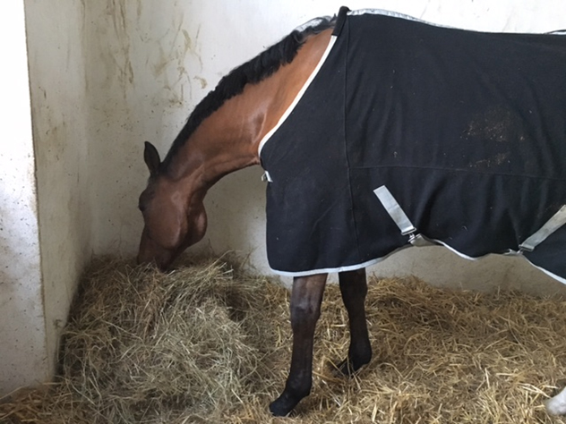
low frequency ultrasound therapy treatment of superficial digital flexor tendon

2nd examination April 28, 2017 (one month later)

  • Longitudinal scan shows straighter SDFT and skin profile
  • Ultrasonographic signs of longer tendon fibers compared to the ones of March 30, 2017
equine chronic tendonitis 5
equine chronic tendonitis 7
equine chronic tendonitis 6
equine chronic tendonitis 8
  • Clinically, RH SDFT appears less swollen and painful
  • Improvement also on ultrasound examination: the anechogenic tendon split is now less visible
  • Horse goes back home with a 4 months rehabilitation program. Same orthopedic shoes

RH - March 30 - 2017

RH - April 28 - 2017

  • Re-check every month
  • Good clinical and US improvement
  • Decrease size of SDFT
  • Painless at palpation
  • Progressive increased level of training
  • 1st competition: August 26 2017 (2nd place) only 5 months after RH SDFT tendinopathy recurrence

Ultrasound examination December 1, 2017
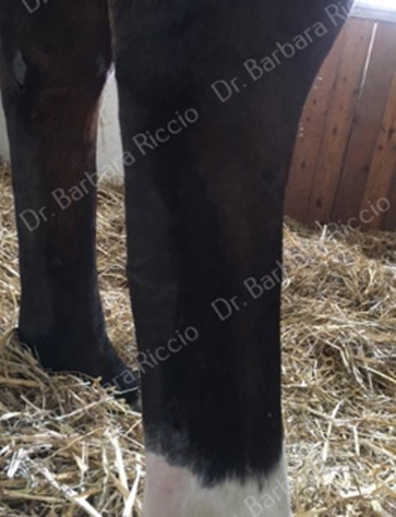
RH - December 1 - 2017

November 2018

  • Between August 2017 and October 2018 horse participated at 40 high level dressage competitions
  • Always made podium
  • 2017 JR Italian Champion
  • Follow-up in full training = 14 months
  • Good outcome: no recurrences!
low frequency ultrasound treatment of SDFT lesion 1
low frequency ultrasound treatment of SDFT lesion 2

Add a comment

*Please complete all fields correctly